肺癌放射性肺炎危险因素的Meta分析
段晨阳a, 刘梦颖a, 吴剑a, 张杰a, 张吉强b
解放军第三军医大学 a. 临床医学系,b. 基础部神经生物学教研室, 重庆 400038
通讯作者:张吉强,Tel: 023-68752232; E-mail:zhangjqtmmu@yahoo.com

作者简介:段晨阳与刘梦颖并列第一作者。段晨阳(1991-),男,石家庄人,在读硕士研究生,主要研究方向为肿瘤医学研究。刘梦颖(1992-),女,河北沧州人,在读硕士研究生,主要研究方向为肿瘤医学研究。

摘要

目的 探讨影响放射性肺炎发生的因素,为更好地指导临床治疗、减少放射性肺炎的发生提供依据。方法计算机检索PubMed数据库、EMBASE数据库、Cochrane协作网和CNKI数据库,并辅以手工检索等方法,应用RevMan 5.1软件对截止2012年2月发表的预测肺癌放疗后发生放射性肺炎的文献数据进行Meta分析。结果共纳入文献75篇。暴露因素包括患者自身因素(性别、年龄、慢性肺疾病、肺功能、合并糖尿病)、肿瘤部位、治疗方案(放疗前有无手术、是否行联合放化疗及使用放疗增敏剂阿米福汀)。Meta分析结果显示其比值比及其95%可信区间分别为:性别0.97[0.82,1.15],年龄0.90[0.63,1.28],合并慢性肺疾病2.18[1.59,3.00],放疗前肺功能0.27[0.11,0.65],合并糖尿病2.46[1.33,4.58],左下肺部肿瘤0.71[0.57,0.90],放疗前肺部手术0.92[0.67,1.25],放化疗联合1.41[1.17,1.71],使用放疗增敏剂阿米福汀2.38[1.79,3.16]。结论影响放射性肺炎发生的因素有合并慢性肺疾病、放疗前肺功能、糖尿病、左下肺部肿瘤、放化疗联合以及使用放疗增敏剂阿米福汀。本项研究的结果认为肺功能较好且无慢性肺疾病和糖尿病等合并症的上肺肺癌患者,在单纯放疗的基础上加用放疗增敏剂阿米福汀,可以降低患者的放射性肺炎发生率。

关键词: 肺癌; 放射性肺炎; 放射性肺损伤; 危险因素; Meta分析
中图分类号:R734.2 文献标识码:A
Risk Factors on Radiation Pneumonitis: A Meta-Analysis
DUAN Chen-yanga, LIU Meng-yinga, WU Jiana, ZHANG Jiea, ZHANG Ji-qiangb
a. Department of Clinical Medicine
b. Department of Neurobiology, Third Military Medical University, Chongqing 400038, China
Abstract

Objective To study the risk factors of radiation pneumonitis in order to find prognostic parameters and provide reference standard for the best clinical treatment plan.Methods The database of PubMed, EMBASE, Cochrane Library and CNKI were searched from the date of their establishments to Februɑry 2012, and other supplied sources were also retrieved. Meta-analysis on literatures predicting radiation pneumonitis after radiotherapy were conducted by using RevMan 5.1 software.Results A total of 75 studies were included. The exposure factors included the patient's own factors (sex, age, chronic lung disease, pulmonary function, diabetes), tumor site, therapeutic schedule (operation before radiotherapy, combined radiochemotherapy, using radiotherapy sensitization agent Amifostine). Meta-analysis results showed that the OR and 95%CI of each factors were: 0.97[0.82,1.15], 0.90[0.63,1.28], 2.18[1.59,3.00], 0.27[0.11,0.65], 2.46[1.33,4.58], 0.71[0.57,0.90], 0.92[0.67,1.25], 1.41[1.17,1.71], 2.38[1.79,3.16], respectively.Conclusions The risk factors of radiation pneumonitis are chronic lung disease, pulmonary function, diabetes, tumor located in left lower lung, combined radiochemotherapy and using radiotherapy sensitization agent Amifostine. The study results indicate that upper lung cancer patients with good pulmonary function and without comorbidity such as diabetes or chronic pulmonary disease have less chance getting radiation pneumonitis based on the simple radiotherapy added with Amifostine—a radiotherapy sensitizer.

Key words: lung cancer; radiation pneumonitis; radiation lung injury; risk factors; meta-analysis

肺癌是世界上死亡率最高的恶性肿瘤,大约2/3的肺癌患者需要接受放射治疗。随着放射治疗技术的飞速发展,伴随的毒副作用,包括放射性肺损伤(radiation lung injury)也成为一个日益突出的问题。据国外资料统计,急性放射性肺损伤的发生率为5%~36%[ 1, 2],由此造成的呼吸衰竭是放射性肺损伤的主要致死原因之一。放射性肺损伤分为早期急性放射性肺炎(radiation pneumonitis, RP)和晚期放射性肺纤维化(radiation pulmonary fiberosis, RPF),二者是连续的过程,目前临床上尚没有令人满意的治疗措施和有效的预测手段,而放射性肺炎一旦发生,将直接影响到患者的治疗效果和生存质量。因此,对放射性肺损伤的预测已成为医学领域中的一项重要课题。本研究对截止2012年2月发表的预测肺癌放疗后发生放射性肺炎的文献进行Meta分析,以期得到影响放射性肺炎发生的因素,为更好地指导临床治疗、减少放射性肺炎的发生提供依据。

1 资料与方法
1.1 资料来源

以“肺癌(lung cancer)”、“ 放射性肺炎(radiation pneumonitis)”、“ 放射性肺损伤(radiation lung injury)”为关键词在PubMed数据库、EMBASE数据库、Cochrane协作网以及CNKI数据库中进行检索。所有检索均截止至2012年2月,手工检索相关期刊,必要时向有关专家索要相关研究资料。

1.2 文献筛选

纳入标准:①研究对象为经细胞学或病理学确诊为原发性肺癌的首次行肺部放射治疗的患者;②文献中记录了各暴露因素发生放射性肺炎及非放射性肺炎的例数、百分比或均数±标准差(mean±SD);③对同一研究组观察不同年限的结果,仅取观察年限最长、最新发表的研究。

排除标准:①无法获得全文的会议摘要;②入组患者同时行胸部其他部位的放疗,或曾在治疗前1年内行胸部放疗;③研究结果为放射性肺纤维化;④评分差的文献;⑤重复发表的文献。

1.3 文献筛选及质量评价

由两人各自独立地完成对入选研究进行资料摘录。将发生放射性肺炎的患者视为放射性肺炎组,未发生放射性肺炎的患者视为非放射性肺炎组。

病例对照研究及队列研究使用Newcastle-Ottawa评分标准[ 3],根据病例的选择、设计和分析的可比性、暴露或结局的确定及评估等三方面进行质量评估;随机对照研究采用CONSORT 2010声明进行评分,共计25项,分别从文章题目及摘要、引言、方法(主要包括试验设计、干预措施、结局指标、随机方法、盲法、统计分析等)、结果(主要包括受试者流程、基线资料、结局及危害等)、讨论(局限性、可推广性)及其他信息进行评价[ 4]

1.4 统计方法

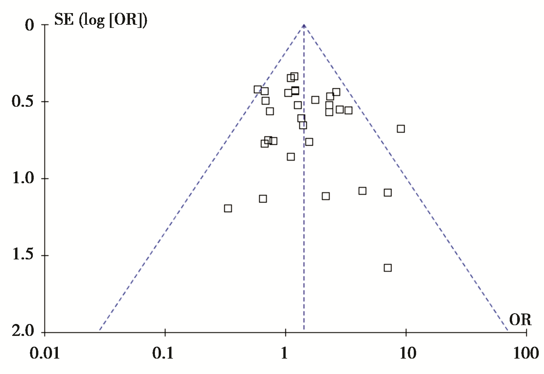
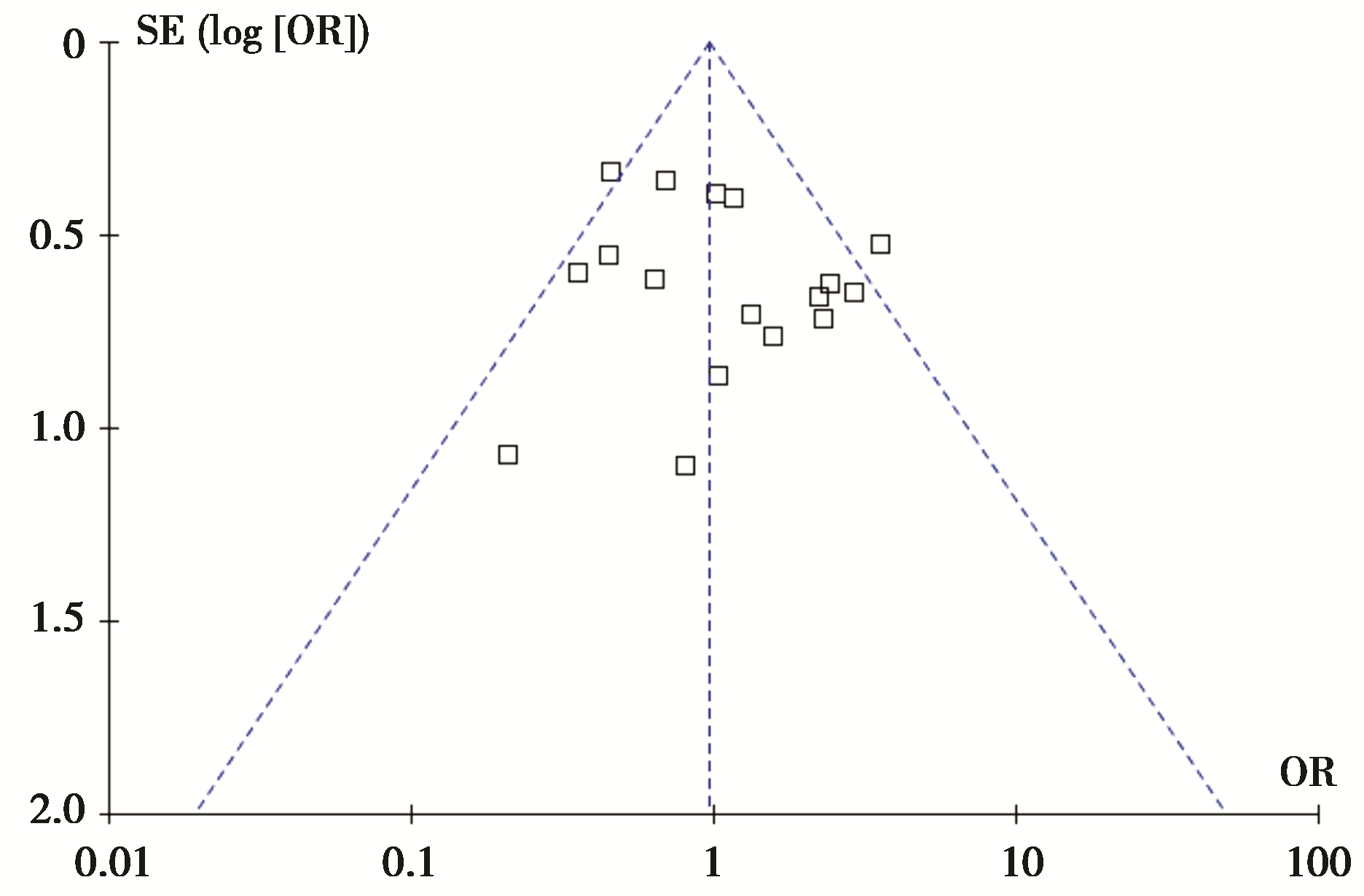
应用Cochrane协作网提供的RevMan 5.1软件。采用Q检验明确有无异质性。经Q检验若无明显统计学差异( P>0.10,或 P≤0.10且 I2≤50%),选用固定效应模型进行分析;若有统计学差异( P≤0.10且 I2>50%),则选用随机效应模型。整合结果以 P≤0.05为差异有统计学意义。敏感性分析用于判断结果的稳定性和强度。主要包括:改变纳入标准、研究质量的高低、统计模型和效应量,观察两种模型的效应值整合结果和异质性有无变化。发表偏倚的识别常采用绘制漏斗图,漏斗图对称则说明发表偏倚得到有效控制。

2 结 果
2.1 文献检索结果

按纳入标准通过浏览目录和摘要初步检索出文献75篇[ 5, 6, 7, 8, 9, 10, 11, 12, 13, 14, 15, 16, 17, 18, 19, 20, 21, 22, 23, 24, 25, 26, 27, 28, 29, 30, 31, 32, 33, 34, 35, 36, 37, 38, 39, 40, 41, 42, 43, 44, 45, 46, 47, 48, 49, 50, 51, 52, 53, 54, 55, 56, 57, 58, 59, 60, 61, 62, 63, 64, 65, 66, 67, 68, 69, 70, 71, 72, 73, 74, 75, 76, 77, 78, 79],其中Nakayama、Vujaskovic等[ 78, 79]的研究因病例数少,根据Newcastle-Ottawa评分较低而排除。纳入的文献中有6篇[ 63, 83, 84, 85, 86, 87]对放射性肺炎的发生率与使用放疗增敏剂阿米福汀的关系进行分析,均为随机对照试验,其中1篇文献有两组数据[ 73]。其余68篇文献均为病例对照研究。纳入的研究均描述了纳入及排除标准,放射性肺炎的诊断标准、随访时间及失访率等内容。暴露因素包括患者自身因素(性别、年龄、慢性肺疾病、肺功能、合并糖尿病),肿瘤部位,治疗方案(放疗前有无手术、是否行联合放化疗及使用放疗增敏剂阿米福汀)等。

2.2 资料和数据提取

对入组文献进行全文阅读,提取了以下内容:①文献一般资料(包括作者姓名、发表年份、作者单位等信息);②研究对象资料(研究例数、是否为原发性肺癌或与其他类型肿瘤同时分析等);③观察终点(放射性肺炎的诊断及分级、放射性肺炎发生例数等);④各个暴露因素下放射性肺炎组与非放射性肺炎组的数值或比例;⑤对于等级资料,提取上述参数不同等级情况下两组的病例数或百分比;对于连续性资料,提取两组的例数及上述参数的mean±SD。

2.3统计学结果

2.3.1患者自身因素

2.3.1.1性别18篇文献2 178例样本纳入分析,Q检验无异质性,采用固定效应模型计算比值比(odds ratio,OR)及95%可信区间(confidence interval,CI)为0.97[0.82,1.15], P=0.73,提示差异无统计学意义。

2.3.1.2年龄10篇文献880例以≤60岁vs. >60岁分析,10篇1 396例以≤70岁vs. >70岁分析, 6篇621例分析年龄mean±SD与放射性肺炎发生率的关系。效应值合并后均无异质性,采用固定效应模型得OR及95%CI值分别为0.90[0.63,1.28]、1.13[0.84,1.52]及-0.01[-1.98,1.96], P分别为0.56、0.34及1.00,均与放射性肺炎的发生无相关性。

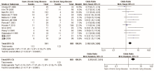
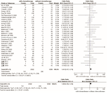
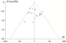
2.3.1.3合并慢性肺疾病13篇共1 409例进行分析,无异质性,固定效应模型得OR及95%CI值为2.18[1.59,3.00], P<0.000 01,提示合并慢性肺疾病的患者容易发生放射性肺炎;调整模型后OR及95%CI值为2.35[1.57,3.51], P<0.000 1,两种模型结果一致,结果可信,见 图1 图2

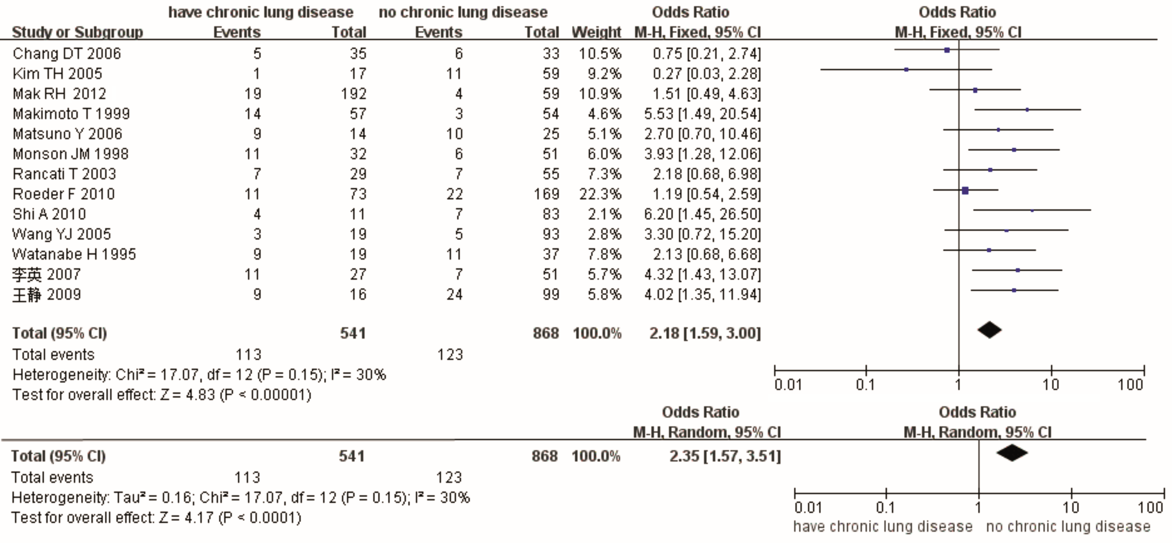
图1 有无慢性肺疾病与RP的效应值比较及敏感性分析图

图2 有无慢性肺疾病与RP的倒漏斗图

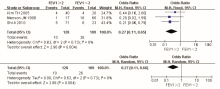
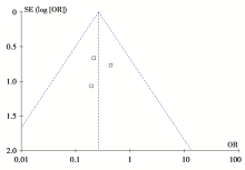
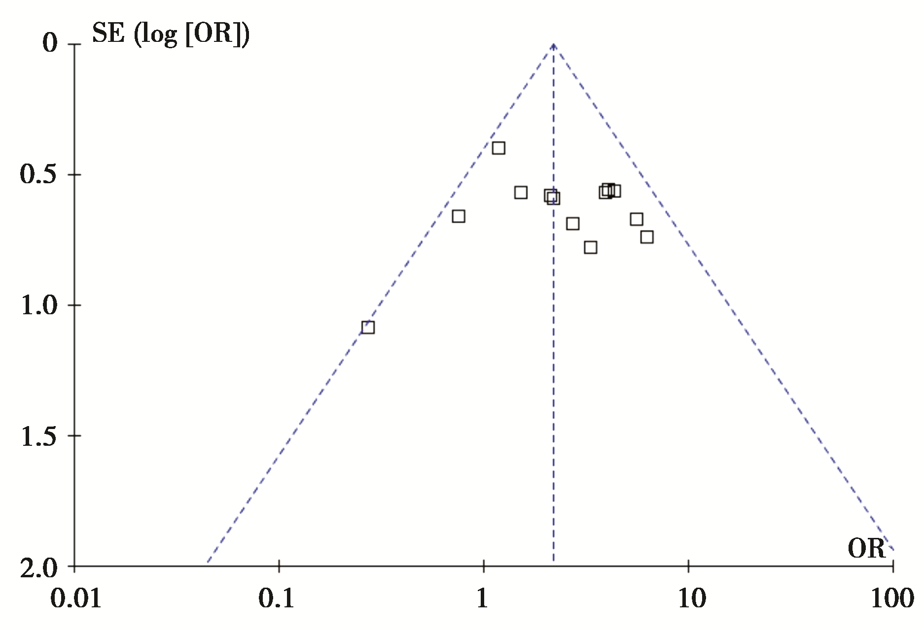
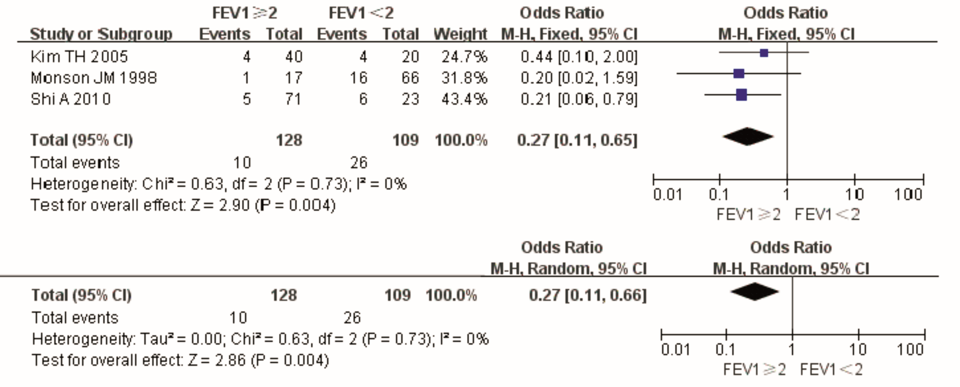
2.3.1.4放疗前肺功能放疗前1秒用力呼气量(forced expiratory volume in 1s, FEV1)≥2L vs. <2L与放射性肺炎的分析,无异质性,固定效应模型得OR及95%CI为0.27[0.11,0.65], P=0.004,提示放疗前FEV1<2L的患者发生放射性肺炎的概率较高。调整模型后,OR及95%CI分别为0.27[0.11,0.66], P=0.004,两种模型结果一致,结果可信,见 图3。但倒漏斗图未完全显示,考虑存在发表偏倚,见 图4

图3 放疗前FEV1≥2或<2L与RP的效应值比较及敏感性分析图

图4 放疗前FEV1≥2或<2L与RP的倒漏斗图

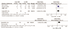
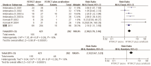
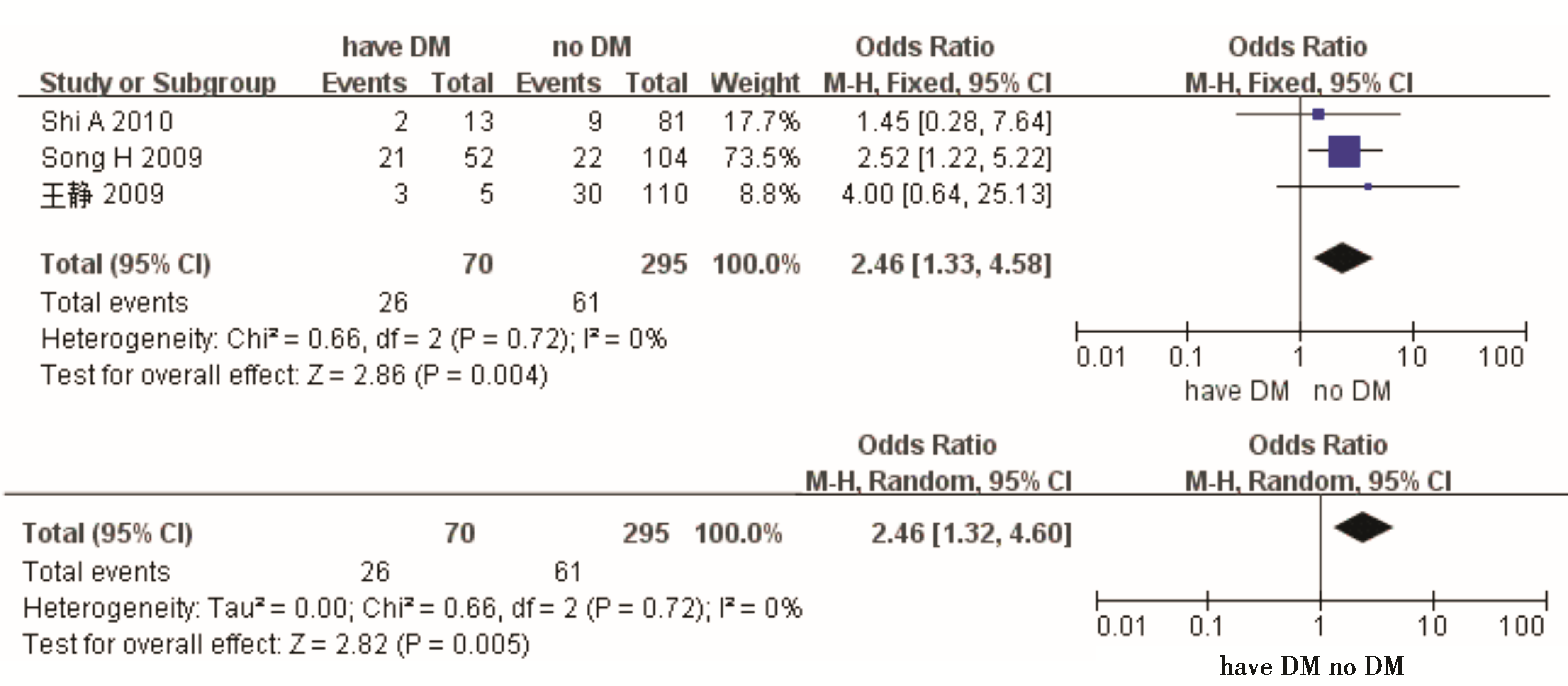
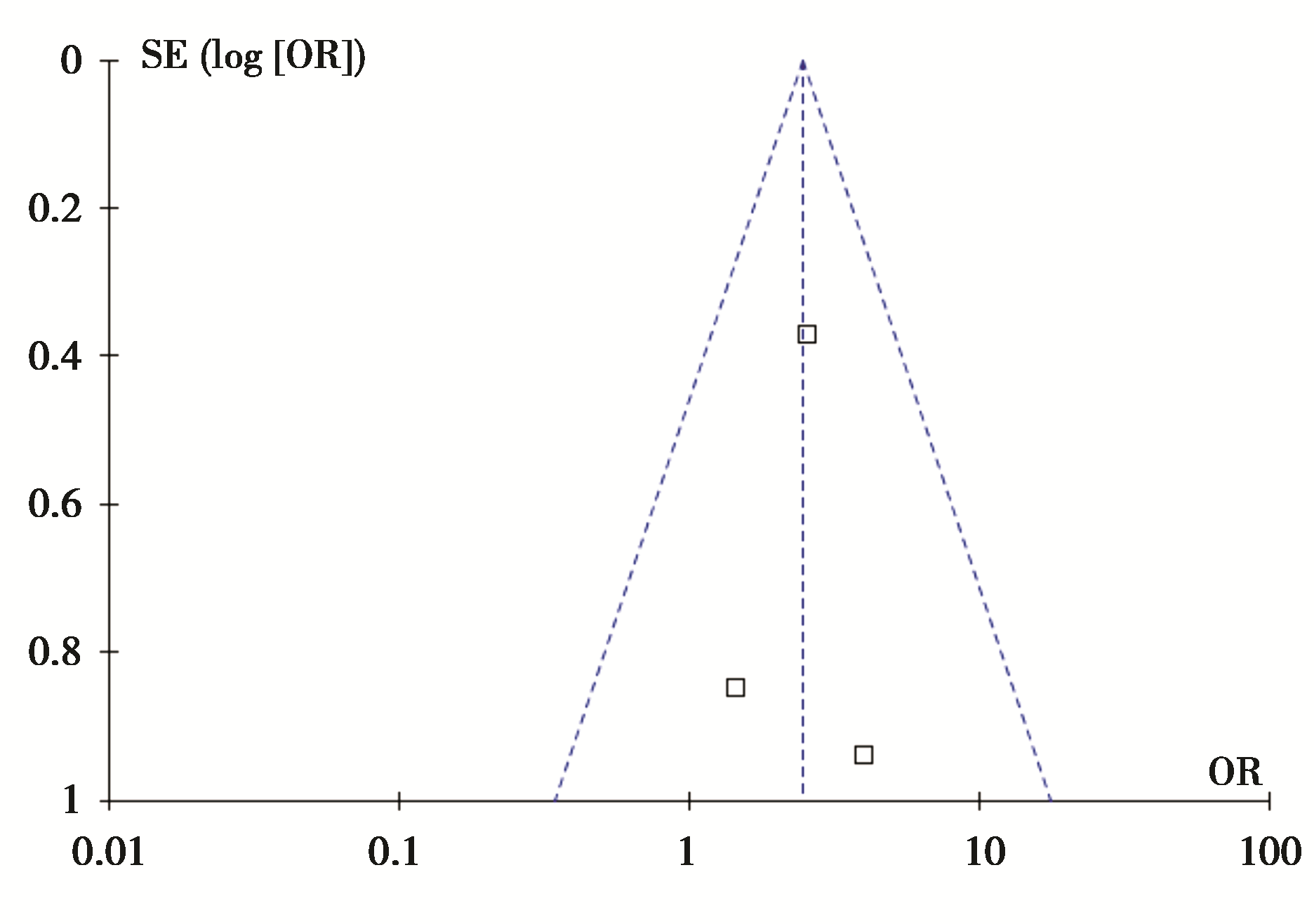
2.3.1.5糖尿病3篇365例样本量对此进行分析,无异质性,OR及95%CI为2.46 [1.33, 4.58], P=0.004,表明合并糖尿病的患者易于发生放射性肺炎。调整模型后,OR及95%CI为2.46[1.32,4.60], P=0.005,两模型结果一致,敏感性较低,稳定性结果,结果可信,见 图5 图6

图5 有无糖尿病与RP的效应值比较及敏感性分析图

图6 有无糖尿病与RP的倒漏斗图

2.3.2肿瘤部位13篇2 211例、9篇1 880例及3篇381例患者分别对上肺vs. 中下肺、右肺vs. 左肺、中央型vs. 周围型进行分析,合并后均无异质性,OR及95%CI分别为0.71[0.57,0.90]、1.18[0.91,1.52]、1.33[0.76,2.35], P为0.005、0.21、0.32,提示肿瘤位于左下肺放疗后易于发生放射性肺炎。调整为随机效应模型,OR及95%CI分别为0.74[0.56,0.99]、 1.17[0.90,1.52]、 1.32[0.74,2.33], P分别为0.04、0.23、0.34,两种结果一致,敏感性较低,稳定性较高,结果可信。

2.3.3治疗因素

2.3.3.1放疗前肺部手术Q检验无异质性,OR及95%CI值为0.92[0.67,1.25], P=0.59,提示无相关性;随机效应模型得OR及95%CI值为0.86[0.53,1.41], P=0.56,两组结果一致,稳定性高,结果可信。

2.3.3.2放化疗联合分别有31篇3 572例和18篇1 760例样本量进行放化疗联合vs.单独放疗、序贯vs. 同步放化疗的分析,均无异质性,OR及95%CI分别为1.41[1.17,1.71]及0.97 [0.76,1.25], P分别为0.000 3及0.83,说明放化疗联合的患者易于发生放射性肺炎;随机效应模型下OR及95%CI分别为1.38[1.10,1.72]及1.02[0.71,1.47], P分别为0.005及0.90,两组结果差别不大,稳定性较高,结果可信,见 图7~ 图10

图7 放化疗联合或单独放疗与RP的效应值比较及敏感性分析图

图8 序贯或同步放化疗与RP的效应值比较及敏感性分析图

图9 放化疗联合或单独放疗与RP的倒漏斗图

图10 序贯或同步放化疗与RP的倒漏斗图

2.3.3.3放疗增敏剂阿米福汀7篇703例样本量进行分析,Q检验无异质性,由于纳入文献为随机对照试验,选用相对危险度(relative risk,RR)作为效应值,固定效应模型得RR及95%CI为2.38[1.79,3.16], P<0.000 01,说明该因素与放射性肺炎的发生有关;调整模型后,RR及95%CI为2.32[1.67,3.23], P<0.000 01,两种结果一致,结果可信,见 图11 图12

图11 单独放化疗或联合阿米福汀与RP的效应值比较及敏感性分析图

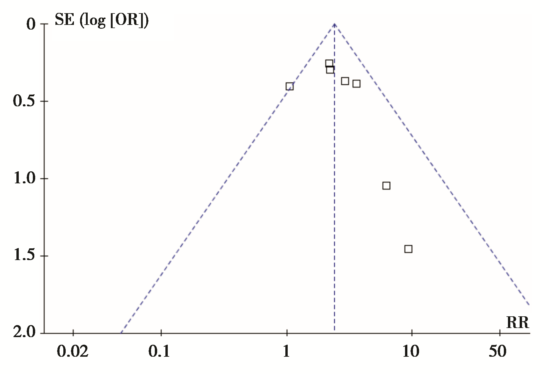
图12 单独放化疗或联合阿米福汀与RP的倒漏斗图

3 讨 论

放射性肺炎是胸部放疗剂量的主要限制性因素,同时也是影响患者放疗后生存质量的最常见因素之一,使得最大程度提高靶区剂量且尽可能减少放射性肺炎的发生成为肺癌治疗策略必须考虑的因素,也是待解决的热点问题之一。

目前部分研究认为,临床因素方面,性别、年龄身体状况及肺功能、肿瘤及治疗因素与放射性肺炎有关。Dang 等[ 11]的研究认为,男性患者放疗后容易发生放射性肺炎,但较多研究认为女性容易发生[ 5, 28],考虑女性肺体积相对较小,同样的照射野更容易发生放射性肺炎。目前的研究普遍认为放化疗联合治疗[ 10, 11, 26, 72]、合并慢性肺疾病[ 7, 16, 26]和放疗前肺功能较差[ 7, 16, 28]的患者存在化疗对肺组织的毒副作用、有效肺体积较小及低氧等引起放射性肺损伤的病理生理过程,因此容易发生放射性肺炎。对于与吸烟的关系,尚未得到统一的结论。Vogelius等[ 80]的系统评价显示老年患者、肿瘤位于中下肺、存在合并症等是放射性肺炎的危险因素,而当前吸烟和有吸烟史是保护因素,考虑可能是吸烟所致的低氧和免疫抑制作用使吸烟患者的肺的耐受性增加。

本文的结果均经过异质性分析及敏感性检验,结果可信。本文还利用敏感性分析评估了发表偏倚对最后结果的可能影响。基于本项研究的结果,我们认为肺功能较好的且无慢性肺疾病和糖尿病等合并症的上肺肺癌患者,在单纯放疗的基础上加用放疗增敏剂阿米福汀,可以降低患者的放射性肺炎发生率。

The authors have declared that no competing interests exist.

参考文献
[1] Medhora M, Gao F, Fish BL, et al. Dose-modifying factor for captopril for mitigation of radiation injury to normal lung[J]. J Radiat Res, 2012, 53(4): 633-640. [本文引用:1] [JCR: 1.447]
[2] Wang D, Sun J, Zhu J, et al. Functional dosimetric metrics for predicting radiation-induced lung injury in non-small cell lung cancer patients treated with chemoradiotherapy[J]. Radiat Oncol, 2012, 7(1): 69. [本文引用:1] [JCR: 2.107]
[3] Stang A. Critical evaluation of the Newcastle-Ottawa scale for the assessment of the quality of nonrand omized studies in meta-analyses[J]. Eur J Epidemiol, 2010, 25(9): 603-605. [本文引用:1] [JCR: 5.118]
[4] Schulz KF, Altman DG, Moher D. CONSORT 2010 Statement: Updated guidelines for reporting parallel group rand omised trials[J]. J Pharmacol Pharmacother, 2010, 1(2): 100-107. [本文引用:1]
[5] Kong FM, Hayman JA, Griffith KA, et al. Final toxicity results of a radiation-dose escalation study in patients with non-small-cell lung cancer (NSCLC): Predictors for radiationpneumonitis and fibrosis[J]. Int J Radiat Oncol Biol Phys, 2006, 65(4): 1075-1086. [本文引用:1]
[6] Barriger RB, Forquer JA, Brabham JG, et al. A dose-volume analysis of radiation pneumonitis in non-small cell lung cancer patients treated with stereotactic body radiation therapy[J]. Int J Radiat Oncol Biol Phys, 2012, 1(82): 457-462. [本文引用:1]
[7] Shi A, Zhu G, Wu H, et al. Analysis of clinical and dosimetric factors associated with severe acute radiation pneumonitis in patients with locally advanced non-small cell lung cancer treated with concurrent chemotherapy and intensity-modulated radiotherapy[J]. Radiat Oncol, 2010, 5: 35. [本文引用:2] [JCR: 2.107]
[8] Claude L, Pérol D, Ginestet C, et al. A prospective study on radiation pneumonitis following conformal radiation therapy in non-small-cell lung cancer: Clinical and dosimetric factors analysis[J]. Radiother Oncol, 2004, 71(2): 175-181. [本文引用:1] [JCR: 4.52]
[9] Mak RH, Alexand er BM, Asomaning K, et al. A single-nucleotide polymorphism in the MTHFR (methylene tetrahydrofolate reductase) gene is associated with risk of radiation pneumonitis in lung cancer patients treated with thoracic radiation therapy[J]. Cancer, 2012, 118(14): 3654-3665. [本文引用:1] [JCR: 5.201]
[10] Ramella S, Trodella L, Mineo TC, et al. Adding ipsilateral V20 and V30 to conventional dosimetric constraints predicts radiation pneumonitis in stage ⅢA-B NSCLC treated with combined-modality therapy[J]. Int J Radiat Oncol Biol Phys, 2010, 76(1): 110-115. [本文引用:1]
[11] Dang J, Li G, Lu X, et al. Analysis of related factors associated with radiation pneumonitis in patients with locally advanced non-small-cell lung cancer treated with three-dimensional conformal radiotherapy[J]. J Cancer Res Clin Oncol, 2010, 136(8): 1169-1178. [本文引用:1]
[12] Zhang L, Yang M, Bi N, et al. ATM polymorphisms are associated with risk of radiation-induced pneumonitis[J]. Int J Radiat Oncol Biol Phys, 2010, 77(5): 1360-1368. [本文引用:1]
[13] Wang LW, Fu XL, Clough R, et al. Can angiotensin-converting enzyme inhibitors protect against symptomatic radiation pneumonitis[J]. Radiat Res, 2000, 153(4): 405-410. [本文引用:1] [JCR: 2.698]
[14] Fujino M, Shirato H, Onishi H, et al. Characteristics of patients who developed radiation pneumonitis requiring steroid therapy after stereotactic irradiation for lung tumors[J]. Cancer J, 2006, 12(1): 41-46. [本文引用:1]
[15] Parashar B, Edwards A, Mehta R, et al. Chemotherapy significantly increases the risk of radiation pneumonitis in radiation therapy of advanced lung cancer[J]. Am J Clin Oncol, 2011, 34(2): 160-164. [本文引用:1] [JCR: 2.552]
[16] Monson JM, Stark P, Reilly JJ, et al. Clinical radiation pneumonitis and radiographic changes after thoracic radiation therapy for lung carcinoma[J]. Cancer, 1998, 82(5): 842-850. [本文引用:2] [JCR: 5.201]
[17] Das SK, Chen S, Deasy JO, et al. Combining multiple models to generate consensus: Application to radiation-induced pneumonitis prediction[J]. Med Phys, 2008, 35(11): 5098-5109. [本文引用:1] [JCR: 2.911]
[18] Roeder F, Friedrich J, Timke C, et al. Correlation of patient-related factors and dose-volume histogram parameters with the onset of radiation pneumonitis in patients with small cell lung cancer[J]. Strahlenther Onkol, 2010, 186(3): 149-156. [本文引用:1] [JCR: 4.163]
[19] Takahashi H, Imai Y, Fujishima T, et al. Diagnostic significance of surfactant proteins A and D in sera from patients with radiation pneumonitis[J]. Eur Respir J, 2001, 17(3): 481-487. [本文引用:1] [JCR: 6.355]
[20] Uno T, Isobe K, Kawakami H, et al. Dose-volume factors predicting radiation pneumonitis in patients receiving salvage radiotherapy for postlobectomy locoregional recurrent non-small-cell lung cancer[J]. Int J Clin Oncol, 2006, 11(1): 55-59. [本文引用:1] [JCR: 1.727]
[21] Fay M, Tan A, Fisher R, et al. Dose-volume histogram analysis as predictor of radiation pneumonitis in primary lung cancer patients treated with radiotherapy[J]. Int J Radiat Oncol Biol Phys, 2005, 61(5): 1355-1363. [本文引用:1]
[22] Kim TH, Cho KH, Pyo HR, et al. Dose-volumetric parameters for predicting severe radiation pneumonitis after three-dimensional conformal radiation therapy for lung cancer[J]. Radiology, 2005, 235(1): 208-215. [本文引用:1] [JCR: 6.339]
[23] Koto M, Tsujii H, Yamamoto N, et al. Dosimetric factors used for thoracic X-ray radiotherapy are not predictive of the occurrence of radiation pneumonitis after carbon-ion radiotherapy[J]. Tohoku J Exp Med, 2007, 213(2): 149-156. [本文引用:1] [JCR: 1.367]
[24] Song H, Yu JM. Effect of diabetes mellitus on the development of radiation pneumonitis in patients with non-small cell lung cancer[J]. Zhonghua Zhong Liu Za Zhi, 2009, 31(1): 45-47. [本文引用:1]
[25] Xiao C, Ding HJ, Feng LC, et al. Efficacy of Liangxue Jiedu Huoxue decoction in prevention of radiation pneumonitis: A rand omized controlled trial[J]. Zhong Xi Yi Jie He Xue Bao, 2010, 8(7): 624-628. [本文引用:1]
[26] Rancati T, Ceresoli GL, Gagliardi G. Factors predicting radiation pneumonitis in lung cancer patients: A retrospective study[J]. Radiother Oncol, 2003, 67(3): 275-283. [本文引用:2] [JCR: 4.52]
[27] Wang YJ, Wang LH, Feng QF, et al. Factors predicting radiation toxicity in the treatment of three-dimensional conformal radiotherapy for lung cancer[J]. Zhongguo Fei Ai Za Zhi, 2005, 8(5): 454-458. [本文引用:1]
[28] Robnett TJ, Machtay M, Vines EF, et al. Factors predicting severe radiation pneumonitis in patients receiving definitive chemoradiation for lung cancer[J]. Int J Radiat Oncol Biol Phys, 2000, 48(1): 89-94. [本文引用:2]
[29] An JY, Kwon SJ, Lee YS. Factors predicting the development of radiation pneumonitis in the patients receiving radiation therapy for lung cancer[J]. Tuberc Respir Dis, 2004, 56(1): 40-50. [本文引用:1]
[30] Hildebrand t MA, Komaki R, Liao Z, et al. Genetic variants in inflammation-related genes are associated with radiation-induced toxicity following treatment for non-small cell lung cancer[J]. PLoS One, 2010, 5(8): e12402. [本文引用:1] [JCR: 3.73]
[31] Huang EX, Hope AJ, Lindsay PE, et al. Heart irradiation as a risk factor for radiation pneumonitis[J]. Acta Oncol, 2011, 50(1): 51-60. [本文引用:1] [JCR: 2.867]
[32] De Ruysscher D, Wand ers R, van Haren E, et al. HI-CHART: A phase Ⅰ/Ⅱ study on the feasibility of high-dose continuous hyperfractionated accelerated radiotherapy in patients with inoperable non-small-cell lung cancer[J]. Int J Radiat Oncol Biol Phys, 2008, 71(1): 132-138. [本文引用:1]
[33] Wang WH, Bao Y, Chen M, et al. Initial outcome of induction chemotherapy with weekly paclitaxel followed by three-dimensional conformal radiotherapy and concurrent weekly paclitaxel for stage Ⅲ non-small cell lung cancer[J]. Ai Zheng, 2006, 25(10): 1279-1283. [本文引用:1]
[34] Vinogradskiy Y, Tucker SL, Liao Z, et al. Investigation of the relationship between gross tumor volume location and pneumonitis rates using a large clinical database of non-small-cell lung cancer patients[J]. Int J Radiat Oncol Biol Phys, 2012, 82(5): 1650-1658. [本文引用:1]
[35] Hope AJ, Lindsay PE, El Naqa I, et al. Modeling radiation pneumonitis risk with clinical, dosimetric, and spatial parameters[J]. Int J Radiat Oncol Biol Phys, 2006, 65(1): 112-124. [本文引用:1]
[36] Lee SW, Choi EK, Lee JS, et al. Phase II study of three-dimensional conformal radiotherapy and concurrent Mitomycin-C, Vinblastine, and Cisplatin chemotherapy for Stage Ⅲ locally advanced, unresectable, non-small-cell lung cancer[J]. Int J Radiat Oncol Biol Phys, 2003, 56(4): 996-1004. [本文引用:1]
[37] Yin M, Liao Z, Huang YJ, et al. Polymorphisms of homologous recombination genes and clinical outcomes of non-small cell lung cancer patients treated with definitive radiotherapy[J]. PLoS One, 2011, 6(5): e20055. [本文引用:1] [JCR: 3.73]
[38] Piotrowski T, Matecka-Nowak M, Milecki P. Prediction of radiation pneumonitis: Dose-volume histogram analysis in 62 patients with non-small cell lung cancer after three-dimensional conformal radiotherapy[J]. Neoplasma, 2005, 52(1): 56-62. [本文引用:1] [JCR: 1.574]
[39] Oh D, Ahn YC, Park HC, et al. Prediction of radiation pneumonitis following high-dose thoracic radiation therapy by 3 Gy/fraction for non-small cell lung cancer: Analysis of clinical and dosimetric factors[J]. Jpn J Clin Oncol, 2009, 39(3): 151-157. [本文引用:1] [JCR: 1.898]
[40] Kobayashi H, Uno T, Isobe K, et al. Radiation pneumonitis following twice-daily radiotherapy with concurrent Carboplatin and Paclitaxel in patients with stage Ⅲ non-small-cell lung cancer[J]. Jpn J Clin Oncol, 2010, 40(5): 464-469. [本文引用:1] [JCR: 1.898]
[41] Inoue A, Kunitoh H, Sekine I, et al. Radiation pneumonitis in lung cancer patients: A retrospective study of risk factors and the long-term prognosis[J]. Int J Radiat Oncol Biol Phys, 2001, 49(3): 649-655. [本文引用:1]
[42] Yamano M, Ogino H, Shibamoto Y, et al. Relationship between radiation pneumonitis and prognosis in patients with primary lung cancer treated by radiotherapy[J]. Kurume Med J, 2007, 54(3-4): 57-63. [本文引用:1]
[43] Segawa Y, Takigawa N, Kataoka M, et al. Risk factors for development of radiation pneumonitis following radiation therapy with or without chemotherapy for lung cancer[J]. Int J Radiat Oncol Biol Phys, 1997, 39(1): 91-98. [本文引用:1]
[44] Yamada M, Kudoh S, Hirata K, et al. Risk factors of pneumonitis following chemoradiotherapy for lung cancer[J]. Eur J Cancer, 1998, 34(1): 71-75. [本文引用:1] [JCR: 5.061]
[45] De Petris L, Lax I, Sirzén F, et al. Role of gross tumor volume on outcome and of dose parameters on toxicity of patients undergoing chemoradiotherapy for locally advanced non-small cell lung cancer[J]. Med Oncol, 2005, 22(4): 375-381. [本文引用:1] [JCR: 2.147]
[46] Makimoto T, Tsuchiya S, Hayakawa K, et al. Risk factors for severe radiation pneumonitis in lung cancer[J]. Jpn J Clin Oncol, 1999, 29(4): 192-197. [本文引用:1] [JCR: 1.898]
[47] Matsuno Y, Satoh H, Ishikawa H, et al. Simultaneous measurements of KL-6 and SP-D in patients undergoing thoracic radiotherapy[J]. Med Oncol, 2006, 23(1): 75-82. [本文引用:1] [JCR: 2.147]
[48] Ishii Y, Kitamura S. Soluble intercellular adhesion molecule-1 as an early detection marker for radiation pneumonitis[J]. Eur Respir J, 1999, 13(4): 733-738. [本文引用:1] [JCR: 6.355]
[49] Wang J, Qiao XY, Lu FH, et al. TGF-beta1 in serum and induced sputum for predicting radiation pneumonitis in patients with non-small cell lung cancer after radiotherapy[J]. Chin J Cancer, 2010, 29(3): 325-329. [本文引用:1] [CJCR: 1.393]
[50] Chang DT, Olivier KR, Morris CG, et al. The impact of heterogeneity correction on dosimetric parameters that predict for radiation pneumonitis[J]. Int J Radiat Oncol Biol Phys, 2006, 65(1): 125-131. [本文引用:1]
[51] Mao J, Kocak Z, Zhou S, et al. The impact of induction chemotherapy and the associated tumor response on subsequent radiation-related changes in lung function and tumor response[J]. Int J Radiat Oncol Biol Phys, 2007, 67(5): 1360-1369. [本文引用:1]
[52] Kocak Z, Yu X, Zhou SM, et al. The impact of pre-radiotherapy surgery on radiation-induced lung injury[J]. Clin Oncol (R Coll Radiol), 2005, 17(4): 210-216. [本文引用:1]
[53] Schild SE, Stella PJ, Geyer SM, et al. The outcome of combined-modality therapy for stage Ⅲ non-small-cell lung cancer in the elderly[J]. J Clin Oncol, 2003, 21(17): 3201-3206. [本文引用:1] [JCR: 18.038]
[54] Clenton SJ, Fisher PM, Conway J, et al. The use of lung dose-volume histograms in predicting post-radiation pneumonitis after non-conventionally fractionated radiotherapy for thoracic carcinoma[J]. Clin Oncol (R Coll Radiol), 2005, 17(8): 599-603. [本文引用:1]
[55] Novakova-Jiresova A, Van Gameren MM, Coppes RP, et al. Transforming growth factor-beta plasma dynamics and post-irradiation lung injury in lung cancer patients[J]. Radiother Oncol, 2004, 71(2): 183-189. [本文引用:1] [JCR: 4.52]
[56] 王静, 乔学英, 曹彦坤, . 非小细胞肺癌三维适形放疗放射性肺炎发生的多因素分析[J]. 中国肿瘤临床, 2009, 36(19): 1086-1089. [本文引用:1]
[57] Watanabe H, Suga A, Tsuchihashi Y, et al. Clinical study of radiation pneumonitis over 10 years[J]. Nihon Kyobu Shikkan Gakkai Zasshi, 1995, 33(4): 384-388. [本文引用:1]
[58] 谢松喜, 李伟雄, 林映如, . 大分割三维适形放疗在非小细胞肺癌治疗中的应用[J]. 中国癌症杂志, 2006, 16(12): 1034-1037. [本文引用:1]
[59] 张彬, 乔田奎. 肺癌同期放化治疗中放射性肺损伤的相关因素分析[J]. 中国癌症杂志, 2010, 37(5): 578-581. [本文引用:1]
[60] 李英, 祝淑钗, 迟子锋. 三维适形放疗肺癌患者的放射性肺炎的相关因素分析[J]. 肿瘤防治研究, 2007, 34(8): 586-589. [本文引用:1]
[61] 钟军, 刘珺, 陈文学, . 血浆TGF-β1与放射性肺损伤的相关性研究[J]. 实用癌症杂志, 2007, 22(5): 468-471. [本文引用:1]
[62] 余娴, 杨镇洲, 王阁, . 62例局部晚期NSCLC三维适形放射治疗中放射性肺炎相关因素分析[J]. 现代肿瘤医学, 2011, 19(6): 1120-1122. [本文引用:1]
[63] 蔡勇, 周道安. 非小细胞肺癌放射性肺炎相关因素分析[J]. 中华医院感染学杂志, 2011, 21(16): 3357-3359. [本文引用:1]
[64] Yorke ED, Jackson A, Rosenzweig KE, et al. Correlation of dosimetric factors and radiation pneumonitis for non-small-cell lung cancer patients in a recently completed dose escalation study[J]. Int J Radiat Oncol Biol Phys, 2005, 63(3): 672-682. [本文引用:1]
[65] Barriger RB, Fakiris AJ, Hanna N, et al. Dose-volume analysis of radiation pneumonitis in non-small-cell lung cancer patients treated with concurrent Cisplatinum and Etoposide with or without consolidation Docetaxel[J]. Int J Radiat Oncol Biol Phys, 2010, 78(5): 1381-1386. [本文引用:1]
[66] Kim JY, Kim YS, Kim YK, et al. The TGF-beta1 dynamics during radiation therapy and its correlation to symptomatic radiation pneumonitis in lung cancer patients[J]. Radiat Oncol, 2009, 4: 59. [本文引用:1] [JCR: 2.107]
[67] Zhang Y, Yu YH, Yu JM. Application of stand ardized uptake value for FDG PET-CT in predicting radiation pneumonitis[J]. Zhonghua Zhong Liu Za Zhi, 2009, 31(8): 622-625. [本文引用:1]
[68] Zhao L, Wang L, Ji W, et al. Association between plasma angiotensin-converting enzyme level and radiation pneumonitis[J]. Cytokine, 2007, 37(1): 71-75. [本文引用:1] [JCR: 2.518]
[69] Evans ES, Kocak Z, Zhou SM, et al. Does transforming growth factor-beta1 predict for radiation-induced pneumonitis in patients treated for lung cancer?[J]Cytokine, 2006, 35(3-4): 186-192. [本文引用:1] [JCR: 2.518]
[70] Kong FM, Anscher MS, Sporn TA, et al. Loss of heterozygosity at the mannose 6-phosphate insulin-like growth factor 2 receptor (M6P/IGF2R) locus predisposes patients to radiation-induced lung injury[J]. Int J Radiat Oncol Biol Phys, 2001, 49(1): 35-41. [本文引用:1]
[71] Anscher MS, Kong FM, Andrews K, et al. Plasma transforming growth factor beta1 as a predictor of radiation pneumonitis[J]. Int J Radiat Oncol Biol Phys, 1998, 41(5): 1029-1035. [本文引用:1]
[72] Fu XL, Huang H, Bentel G, et al. Predicting the risk of symptomatic radiation-induced lung injury using both the physical and biologic parameters V(30) and transforming growth factor beta[J]. Int J Radiat Oncol Biol Phys, 2001, 50(4): 899-908. [本文引用:1]
[73] Antonadou D, Petridis A, Synodinou M, et al. Amifostine reduces radiochemotherapy-induced toxicities in patients with locally advanced non-small cell lung cancer[J]. Semin Oncol, 2003, 30(6 Suppl 18): 2-9. [本文引用:1] [JCR: 4.327]
[74] Antonadou D. Radiotherapy or chemotherapy followed by radiotherapy with or without Amifostine in locally advanced lung cancer[J]. Semin Radiat Oncol, 2002, 12(1 Suppl 1): 50-58. [本文引用:1] [JCR: 3.971]
[75] Komaki R, Lee JS, Kaplan B, et al. Rand omized phase Ⅲ study of chemoradiation with or without Amifostine for patients with favorable performance status inoperable stage Ⅱ-Ⅲ non-small cell lung cancer: Preliminary results[J]. Semin Radiat Oncol, 2002, 12(1 Suppl 1): 46-49. [本文引用:1] [JCR: 3.971]
[76] Antonadou D, Coliarakis N, Synodinou M, et al. Rand omized phase Ⅲ trial of radiation treatment +/- Amifostine in patients with advanced-stage lung cancer[J]. Int J Radiat Oncol Biol Phys, 2001, 51(4): 915-922. [本文引用:1]
[77] Komaki R, Lee JS, Milas L, et al. Effects of Amifostine on acute toxicity from concurrent chemotherapy and radiotherapy for inoperable non-small cell lung cancer: Report of a rand omized comparative trial[J]. Int J Radiat Oncol Biol Phys, 2004, 58(5): 1369-1377. [本文引用:1]
[78] Nakayama Y, Makino S, Fukuda Y, et al. Activation of lavage lymphocytes in lung injuries caused by radiotherapy for lung cancer[J]. Int J Radiat Oncol Biol Phys, 1996, 34(2): 459-467. [本文引用:1]
[79] Vujaskovic Z, Groen HJ. TGF-beta, radiation-induced pulmonary injury and lung cancer[J]. Int J Radiat Biol, 2000, 76(4): 511-516. [本文引用:1] [JCR: 1.895]
[80] Vogelius IS, Bentzen S. Clinical factors associated with risk of radiation pneumonitis: A litterature based meta-analysis[J]. Radiother Oncol, 2010, 96: S125-S126. [本文引用:1] [JCR: 4.52]